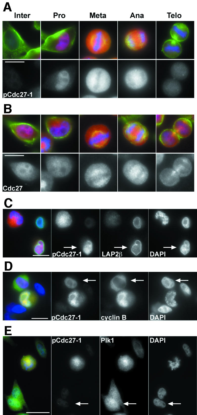
Fig. 5. APC phospho-epitopes appear in prophase when cyclin B1 translocates into the nucleus. (A) HeLa cells were stained with pCdc27-1 antibodies (red), tubulin (green) and DAPI (blue). (B) Staining was performed as in (A) but using non-phospho-specific Cdc27 antibodies (red). (C) HeLa cells treated as in (A) were stained with pCdc27, LAP2β (green) and DAPI (blue). The arrow indicates a nucleus that stains with pCdc27-1 antibodies and is still surrounded by an intact lamina. (D and E) Co-staining of pCdc27-1 with cyclin B1 (D) and Plk1 (E). The arrow in (D) indicates a cell where nuclear uptake of cyclin B1 has just begun and where pCdc27-1 staining can be seen in the nucleus. The arrow in (E) indicates a cell in which Plk1 but no pCdc27-1 staining can be detected in the nucleus. Bar, 10 µm (A and B), 20 µm (C–E).
