
Figure 2.
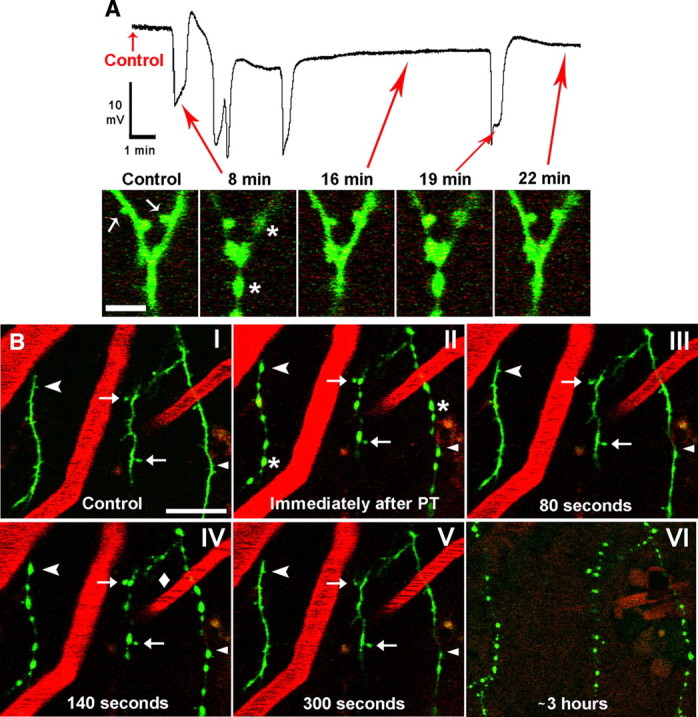
Dendrites in the penumbra undergo a rapid cycle of beading and recovery coinciding with the passage of SDs. A, Top: example recording from microelectrode placed next to imaged dendrites within the middle of penumbra, showing the photothrombotic occlusion-induced initial SD (∼8 min) followed by 4 spontaneous SDs. Note a small DC negative shift on which spontaneous SDs are riding. Bottom, In vivo 2PLSM high-magnification image sequence showing a branched dendrite from layer I of somatosensory cortex with arrows corresponding to various time points on the above physiology recording. The dendrite (spines indicated by arrows) appears normal in control but becomes beaded (asterisks) and spines are lost immediately after induced SD at 8 min. The dendrite and spines are shown to be recovered at 16 min. However, a subsequent spontaneous SD at 19 min beads the dendrite again, with recovery shown at 22 min. Scale bar, 5 μm. B, Low-magnification 2PLSM image sequence showing dendrites (green) as well as blood vessels (red; labeled with Texas Red dextran) from layer I of somatosensory cortex. Blood flow within vessels is indicated by streaking caused by scanning of moving nonfluorescent red blood cells. The dendrites appear normal during control (I) and undergo rapid beading (asterisks) and recovery coinciding with SDs (II–V) in similar fashion to the example shown in A. Blood flow velocity in one of the vessels slows during an SD (IV, diamond) as dendrites bead, despite no apparent change in the other two imaged vessels. Some dendritic spines are permanently lost (arrowhead), transiently lost (chevrons), or persistent (arrows) during SD-induced dendritic beading. Appearance of dendritic beads is relatively constant during passage of these SDs. Finally, nearly 3 h after photothrombosis, the dendrites undergo terminal beading after an SD and are no longer able to recover (VI) (vessels are no longer clearly seen at this time point due to Texas Red Dextran leaking out, necessitating a reduction in the red channel gain). Scale bar, 20 μm.