Figure 2.
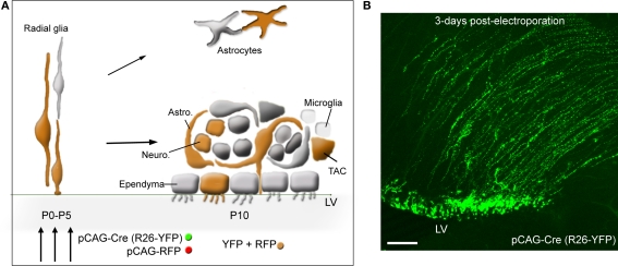
SVZ electroporation and labeled cells. (A) Diagram illustrating: (1) the transformation of radial glia into SVZ astrocytes and ependymal cells, and parenchymal astrocytes during the first 2 weeks, (2) the cellular organization of the SVZ. Astrocyte-like cells (astro.) ensheath neuroblasts (neuro.). TACs and microglia (represented as small light cells) are scattered throughout the SVZ. Ependymal cells line the lateral ventricle (LV). (3) Co-electroporation of pCAG-driven fluorescent proteins results in protein expression into radial glia, and thus in SVZ astrocyte-like cells and ependymal cells, and the progeny of astrocyte-like cells, i.e., TAC and neuroblasts that appear orange due to both RFP and YFP co-localization. The diagram assumes a 100% co-localization, which is experimentally 80–90%. (B) Confocal image illustrating the expression of yellow fluorescent protein (YFP) in radial glia following electroporation of a pCAG-Cre in Rosa26-YFP mice. Scale bar: 100 μm.

