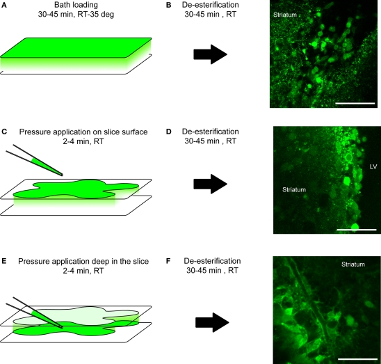
Figure 5.
Loading protocol of calcium indicator dyes. (A, C, E) Diagram illustrating the loading protocol of a calcium indicator dye: by bath also called bulk loading (A), by pressure application on top of the tissue (C) or inside the tissue (E). RT, room temperature; deg, degree. (B, D, F) After a 30- to 45-min de-esterification time-period at room temperature (RT), loading cells progressively become fluorescent upon proper excitation as shown on confocal images on the right. Scale bar: 50 μm (B, D, F).

