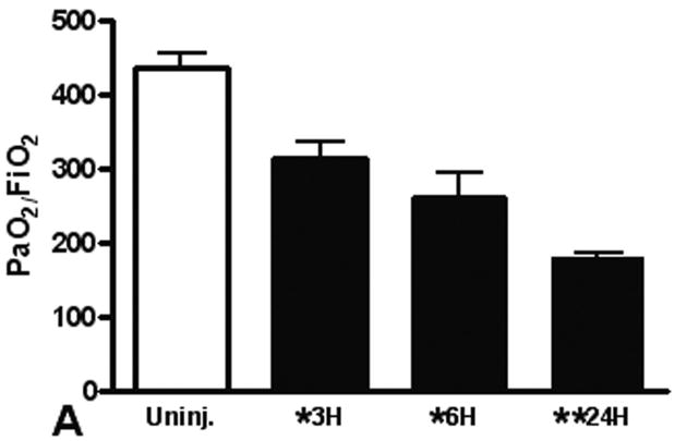
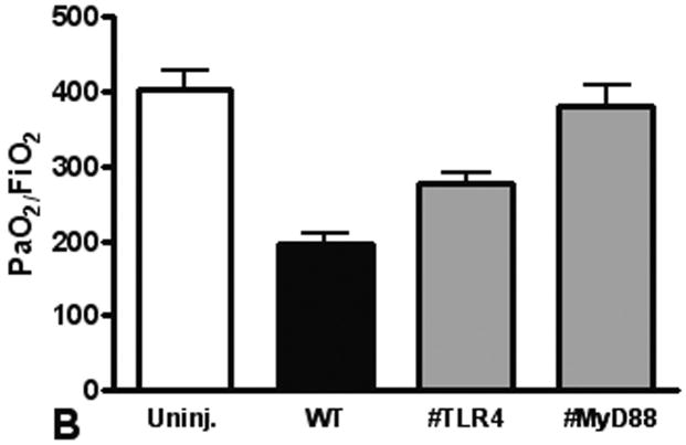
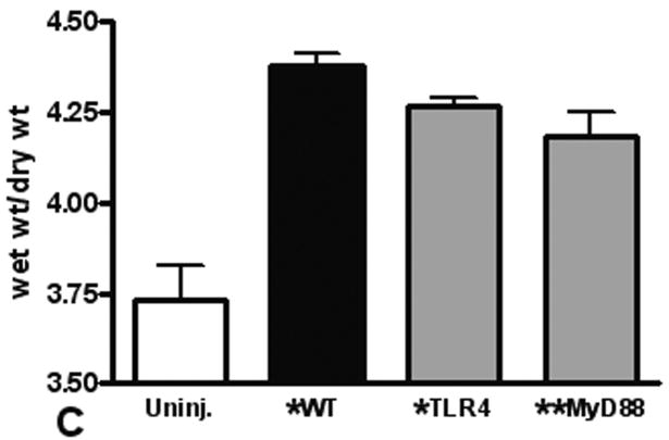
Figure 1. Lung dysfunction after pulmonary contusion is dependent on the TLR4 pathway.
Arterial blood from uninjured (open) and injured animals (closed) was isolated and blood gas analysis performed as described in the methods. (A) PaO2:FiO2 ratios were calculated and are shown for WT animals at 3, 6 and 24 hours after injury (n=5/time). There is a significant decrease in PaO2:FiO2 in WT animals after injury (*p≤0.01), with the most significant decrease at 24H (**p<0.01 relative to other injury times). (B) Blood gas analysis on injured TLR4 and MyD88 deficient animals (gray) at 24H shows significantly increased PaO2:FiO2 ratios when compared to WT injured animals (#p≤0.05, n=7/genotype). (C) WT (black) and TLR4 and MyD88 deficient (gray) injured lungs were isolated at 24H after injury and weighed before (wet wt.) and after drying (dry wt.) as described in the methods (n=5/genotype). Pulmonary edema (wet wt/dry wt) is significant (*p≤.05) in all animals and is significantly decreased in MyD88 deficient mice (**p<.05 compared to WT).


