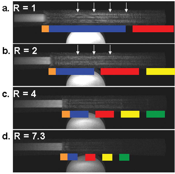
Figure 8.

Selected frames from an EC CAPR acquisitions (bolus velocity 8 mm/s) with varying 2D SENSE accelerations: (a) R=1, (b) R=2, (c) R=4, (d) R=7.3 reconstructed with Delay 3. The k-space sampling pattern is superimposed in color. Motion is left to right. With increased SENSE acceleration there is a decrease in the temporal footprint, decreased anticipation artifact (arrows, a and b), and improved leading edge sharpness.
