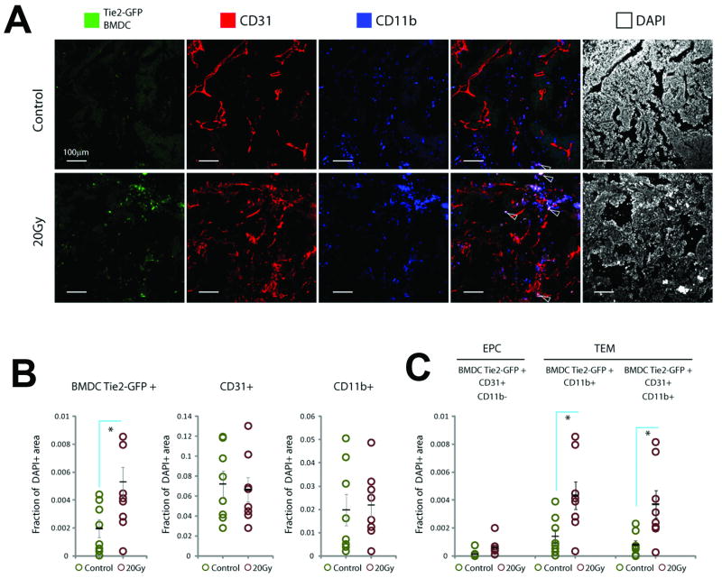
Figure 4. Analysis of BMDCs in tumors recurring after local irradiation in WT/Tie2-GFP-BMT mice.
A, Representative confocal microscopy images of fluorescence immunohistochemistry in tumors recurring after 20Gy of radiation versus non-irradiated size-matched (control) tumors. Note localization of GFP expression (green) in CD11b+ cells outside vessels (blue, see arrows) but not in CD31+CD11b− vascular endothelial cells (red). B,C, Quantification of BMDCs: Overall myeloid cell infiltration (CD11b+) and CD31+ microvascular density were not significantly changed, but the total number of Tie2+ BMDCs increased in irradiated tumors (B). These represented mostly Tie2+CD11b+ TEMs, the majority of which were also CD31+ but had peri-vascular location; in contrast the number of vessel-incorporated Tie2+CD31+CD11b− EPCs was negligible and not different in tumors grwoing after irradiation (C). (*denotes p<0.05).

