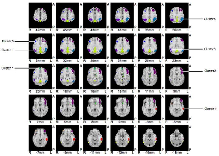
Figure 1.

Cluster division for SVM based on the subtraction of FDG PET scans of healthy controls from all MCI patients after age regression (i.e., MCI template). Note the similarity to AD pattern. Top of image is anterior; image left is patient’s right side. Each cluster has been colorized to aid in identification.
