
Figure 4.
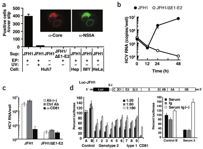
Infectivity of viral particles and neutralization of infection. (a, insert) Immunofluorescence analysis of cells infected with viral particles. Huh7 cells inoculated with virus-containing medium were stained simultaneously for core (left) and NS5A (right). (a) Cell lines specified on the bottom were also inoculated with a 30-fold concentrated supernatant from full-length JFH1 RNA– or JFH1/ΔE1-E2 RNA– transfected cells (Sup). In some experiments, culture supernatant of nontransfected cells was used (EP−), or culture supernatant was irradiated with ultraviolet light before inoculation of cells (UV+). We stained cells 2 d after infection for core protein and counted positive cells. (b) Comparison of infectivity of culture supernatant from JFH1 RNA– and JFH1/ΔE1-E2 RNA– transfected cells. Supernatants containing about 108 RNA copies/ml were used for inoculation of Huh7 cells and amounts of cell-associated RNA were determined 0, 12, 24 and 48 h after inoculation. (c) Inhibition of infection by CD81-specific antibody. We used 20-fold concentrated supernatants from cells transfected with given genomes for infection of Huh7 cells in the presence of a CD81-specific (α-CD81, black bars) or a control antibody (Ctrl Ab, gray bars), or in the absence of antibody (Ab(−), white bars). Inoculated cells were analyzed by RTD-PCR. (d) Production of infectious HCV particles carrying the firefly luciferase reporter gene and neutralization of infection by sera from infected individuals. Upper panel, schematic representation of Luc-JFH1 construct with luciferase (Luc) reporter gene (Supplementary Fig. 6 online). E-I, EMCV-IRES. Bottom left panel, neutralization of Luc-JFH1 virus by sera from infected individuals. Culture supernatants containing Luc-JFH1 viral particles were mixed with given dilutions of sera from healthy donor (Control), or sera from individuals infected with given genotypes (lanes 1–9). Results of CD81-specific antibody neutralization are shown in the right; black bar, 2 μg/ml; gray bar, 0.4 μg/ml; white bar, 0.08 μg/ml. Luciferase activity was determined 72 h later and is expressed relative to the values obtained with control serum A. Bottom right panel, neutralization by immunoglobulins purified from infected individuals' sera. Luc-JFH1 virus–containing supernatant was mixed with 2 mg total protein contained in control serum B or patient serum 3 (serum, black bars). Addition of this amount of serum protein is equivalent to a final serum dilution of approximately 1:20. Alternatively, virus preparation was mixed with 2 mg protein of the same sera depleted of immunoglobulins (Serum Ig(−), open bars), or 130 μg of corresponding purified immunoglobulins (Ig, gray bars). Infectivity is expressed relative to control serum B.