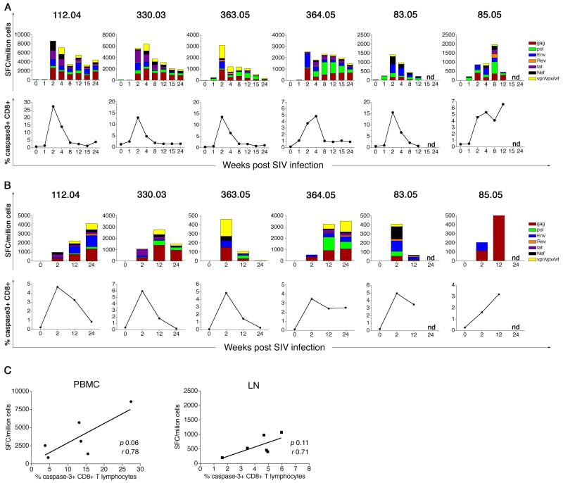
Figure 5. SIV-specific IFN-γ ELISPOT responses and CD8+ T lymphocyte apoptosis in SIVmac239-infected rhesus macaques.
(A) Kinetics of peripheral blood SIV-specific IFN-γ ELISPOT responses (top panel) and CD8+ T lymphocyte apoptosis (bottom panel) for the first 24 weeks following SIV infection. (B) Kinetics of peripheral lymph node SIV-specific IFN-γ ELISPOT responses (top panel) and CD8+ T lymphocyte apoptosis (bottom panel) for the first 24 weeks following SIV infection. Data on individual SIVmac239-infected rhesus macaques shown. (C) Relationship between the magnitude of the SIV-specific IFN-γ ELISPOT response and frequency of active caspase-3-positive CD8+ T lymphocytes at two weeks post SIV infection in PBMC (left panel) and lymph node (right panel). Correlation analysis was performed with the Pearson test. SFC: Spot forming cells

