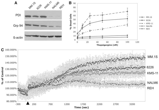
Fig. 1.
Higher expression of ER-resident proteins correlates with hypersensitivity to thapsigargine and increased ER Ca2+ leak in MM as compared to non-myeloma cell lines. a Expression of two ERresident proteins, GRP94 and PDI, was assayed by western blot in all 5 cell lines. b Sensitivity to the SERCA inhibitor, thapsigargine, in MM cell lines, as assayed by trypan blue exclusion assays following 24 h treatment. The data is an average of triplicate samples from one of three experiments. c ER Ca2+ leak was estimated by the increase in the ratio of indo-1 fluorescence emitted at 400 versus 500 nm following thapsigargine treatment. Thapsigargine was added after 5 min of basal calcium measurement. The data represents the percent increase of the ratio of indo-1 fluorescence as compared to control levels and the average + SD of triplicate samples

