Fig. 4.
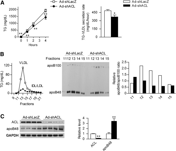
4. Hepatic ACL deficiency suppresses the secretion of apoB48-containing VLDL. A: Male C57BL/6J mice at 8 weeks of age were infected with Ad-shLacZ or Ad-shACL and maintained on LFD for 18 days (n = 8/group). After a 4 h fast, tyloxapol was administered through tail-vein injection at 600 mg/kg to block the LPL activity. Blood triglycerides were measured at the time points as indicated, and the VLDL secretion rate was derived from the slope of the line using least squares regression. Data are shown as means ± SEM. *P < 0.05 and **P < 0.01 versus Ad-shLacZ-infected mice by t-Test. B: Mice likewise infected were treated with tyloxapol for 4 h, and serum samples were collected and pooled for FPLC fractionation analysis (n = 4). The TG concentration was determined from each of the indicated fractions, and equal volumes of the VLDL fractions were subjected to Western immunoblot analysis using the apoB antibody. The bar graph indicates the relative apoB48:apoB100 ratio after quantification by densitometry from the immunoblots. C: Western immunoblot analysis of the liver ACL and apoB48 protein abundance from the mice treated for 4 h with tyloxapol as in (B). Each lane represents analyzed sample from an individual mouse. The bar graphs indicate the relative abundance of ACL and apoB48 protein as quantified by densitometry from the immunoblots after normalization to GAPDH (n = 4/group). Data are shown as means ± SEM. **P < 0.01 versus Ad-shLacZ-infected mice. ACL, ATP-citrate lyase; Apo, apolipoprotein; FPLC, fast-protein liquid chromatography; HFD, high-fat diet; LFD, low-fat diet; TG, triglyceride.

