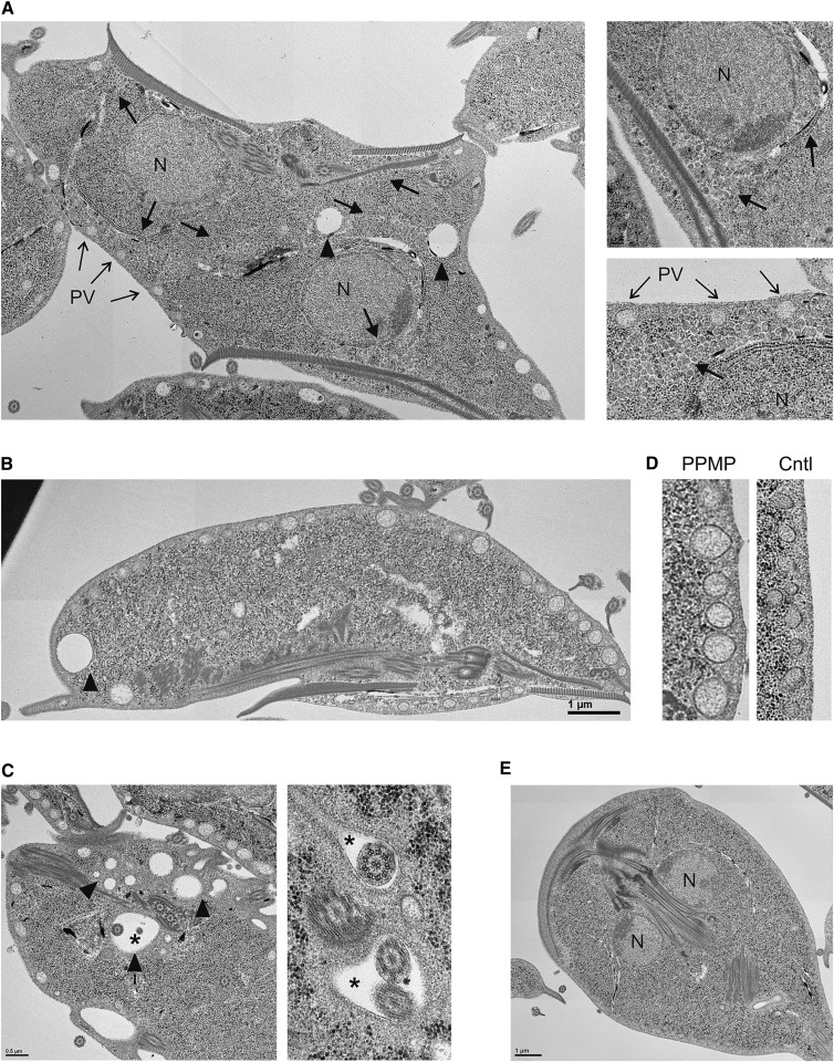
Fig. 6.
Ultrastructural abnormalities following inhibition of GlcCer synthesis. Electron micrographs of G. lamblia treated with 10 μM PPMP for 16 h. Note the cytosolic accumulation of vesicles (A, thick arrows), the electron-lucent vacuoles of various diameters (A, B, C, arrowheads), the presence of vacuoles containing parasite flagella (C, asterisks), and distended peripheral vesicles (D). Untreated control parasites (E). N, nucleus. PV, peripheral vesicles. Scale bar: 3 μm; except in panel D and right part of panel C, 0.5 μm.

