Fig. 7.
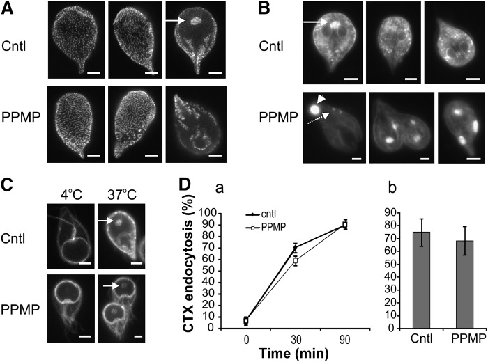
Inhibition of GlcCer synthesis alters clathrin localization and endolysosomal compartments. A: Parasite cultures were treated with solvent (cntl) or 10 μM PPMP for 16 h and stained with anti-clathrin (CLH) antibody. Note the aggregation of CLH-positive punctuated structures upon PPMP treatment. Arrow, signature site for endocytosis. B: Lysotracker™ staining of acidic compartments in parasites treated as in panel A. Note the reduction of the punctuated staining (dashed arrow) and the appearance of large structures (arrowhead) in PPMP-treated cells. Arrow, signature site for endocytosis. C: Parasite cultures were treated with solvent (cntl) or 10 μM PPMP for 16 h and stained with cholera toxin (CTX) at 4°C to label the plasma membrane. Following a 37°C incubation, CTX is internalized and labels the endocytosis signature site (arrow) in both control and drug treated samples. D. (a) Time course of cells treated for 30 min with 10 μM PPMP, stained with CTX at 4°C and showing the endocytosis signature site following a 37°C incubation for the indicated time. (b) Cells were treated with 10 μM PPMP for 16 h, labeled with CTX as before and counted for endocytosis signature site-staining after 90 min of 37°C incubation. Results are presented as percentage of total parasite number ± SE (n = 6) of two experiments done in triplicate. Scale bars: 3 μm.

