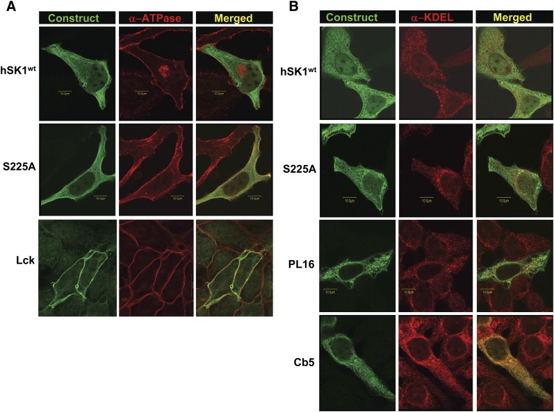
Fig. 1.
Intracellular localization of recombinant hSK1 constructs as assessed by confocal microscopy. HeLa cells were transiently transfected with recombinant hSK1wt; the phosphorylation-deficient mutant hSK1S225A; the plasma membrane-targeted construct Lck; or the endoplasmic reticulum-targeted constructs PL16 and Cb5, as described. Cells were immunostained with α-FLAG antibody (green) for detection of transfected constructs in conjunction with antibodies for specific intracellular markers (red). Immunofluorescence was analyzed by confocal microscopy, and images are presented as representative Z-stacks or individual confocal slices. Merged images (yellow) represent the amount of colocalization of each hSK1 construct with the specified intracellular marker. A: Immunostaining for the plasma membrane marker, the α subunit of ATPase. B: Immunostaining for endoplasmic reticulum as assessed by α-KDEL immunoreactivity. Abbreviations: HeLa, human cervical carcinoma; SK1, sphingosine kinase 1.

