Fig. 5.
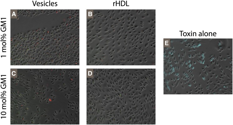
Localization of toxin and decoy after incubation. Fluorescently-labeled lipid particles and cholera toxin B with human retinal pigment epithelial cells. Cells appear in gray with NBD-labeled rHDL in green, Alexa594-CTB in red, and FITC-CTB in cyan (E only). FITC-CTB was used for experiments involving only one fluorophore while Alexa594-CTB was used for experiments requiring multiple fluorophores to allow imaging of each probe on a separate channel. All images are contrast enhanced in a standardized fashion and captured at 10× magnification. CTB, cholera toxin subunit B; FITC, fluorescein-isothiocyanate; GM1, monosialotetrahexosylganglioside.

