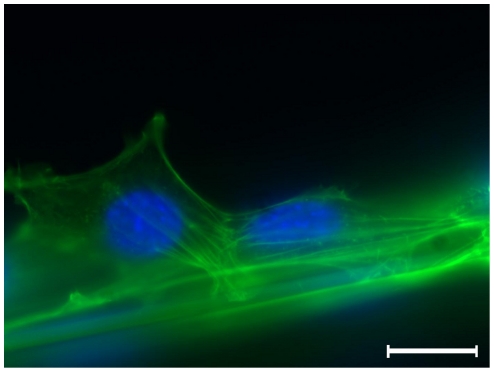
Figure 3. Immunofluorescence microscopy of fibroblasts adhering to a spider silk fibre.
Fibroblasts are sticking to fibre, note the orientation of intracellular actin filament bundles, indicating the direction of forces; DAPI staining of cell nuclei in blue, α-actin as well as autofluorescence of spider silk in green; magnitude ×400, scale bar represents 10 µm.

