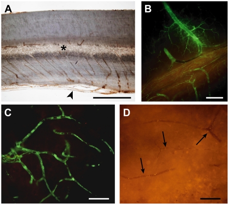
Figure 10. Vascular supply after spinal cord injury.
A) Sagittal section (50 µm) of rat thoracic spinal cord showing the gross vascular anatomy. Note that parallel sulcal arteries, at every 50 to 100 µm along the cord, extending from the anterior spinal artery (arrowhead) into the central grey matter (*). This suggests a segmented vascular supply of the central grey matter. B–D) Sagittal sections (50 µm) of rat thoracic spinal cord 15 min after i.v. injection of a 70 kDa green fluorescent dextran into a rat 4 days after spinal injury. B) In this section from just outside the injury site, vessels are filled with the dextran indicating that blood flow is maintained in these vessels. C) Higher magnification shows that the dextran has reached small capillaries just outside the injury zone. This is in contrast to the injury site where capillaries are devoid of the dextran indicating that flow through these vessels is impeded (arrows in D). The highly segmental arrangement of the blood supply to the central grey matter (A) could account for the limited rostral and caudal expansion of the lesion since the blood supply to regions outside the initial injury zone appears to be maintained (B & C). Scale bars are 1 mm in A, 250 µm in B and 50 µm in C & D.

