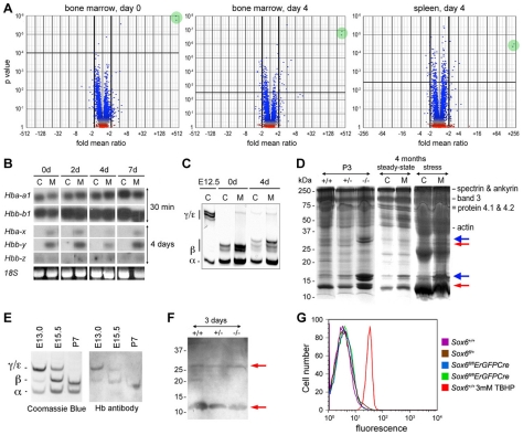
Figure 5. Embryonic globin and other gene expression changes in Sox6-deficient erythropoietic tissues and primary erythroid cells.
(A) Volcano plots showing gene expression changes in mutant tissues. RNA was from bone marrow of Sox6fl/fl and Sox6fl/flErGFPCre mice before PHZ treatment (day 0) [left]; bone marrow [middle] and spleen [right] of similar mice 4 days after treatment. Each dot represents a gene probe. Green circles highlight Hbb-y identified by two probes. (B) Northern blot of globin RNA levels in spleen of Sox6fl/fl (C) and Sox6fl/flErGFPCre (M) mice 0 to 7 days after PHZ treatment. Probes were for adult alpha-globin (Hba-a1) and beta-globin (Hbb-b1), and embryonic X-globin (Hba-x), Y-globin (Hbb-y), and Z-globin (Hbb-z). All had similar specific activity and length. X-ray films were exposed for 30 min for adult globin RNAs, and 4 days for embryonic globin RNAs. The latter were thus weakly expressed compared to the former. (C) Electrophoretic analysis of globins in RBCs from E12.5 wild-type embryo and control and mutant adult mice 0 and 4 days after PHZ treatment. Globin chains are indicated. Several beta-globin isoforms are seen due to mixed genetic background. (D) RBC ghosts from 3-day-old (P3) Sox6+/+, Sox6+/−, and Sox6−/− littermates, and 4-month-old Sox6fl/fl (C) and Sox6fl/flErGFPCre (M) mice 0 day (steady-state) and 4 days (stress erythropoiesis) after PHZ treatment. Protein marker molecular weights and typical RBC proteins are indicated. Blue arrows, ectopic histones in mutants. Red arrows, proteins reacting with hemoglobin antibody against beta-, gamma-, delta-, and epsilon-globins. (E) Coomassie blue staining of globins in RBC lysates from control mice at E13.0, E15.5, and postnatal day 7 (P7) and western blot with hemoglobin antibody. (F) Western blot of ghosts from P3 mice with hemoglobin antibody. Red arrows, all samples have similar residual globin chains. (G) Flow cytometry shows that minimal ROS production in blood of control and mutant adult mice, except in control blood treated with tert-butyl hydroperoxide (TBHP).

