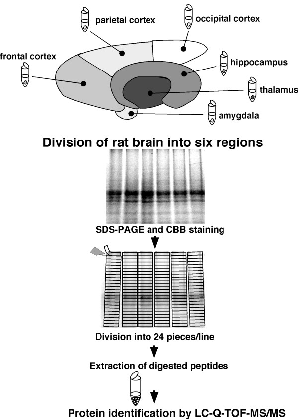
Figure 1.
Flow diagram of the experimental design. Rat brains were divided into six regions: thalamus, hippocampus, frontal cortex, parietal cortex, occipital cortex, and amygdala. The divided samples were lysed in lysis buffer containing SDS, and subjected to SDS-PAGE with Coomassie Brilliant Blue staining. The gel lane was divided into 24 slices, and the slices were pre-treated by in-gel trypsin digestion. The amino acid sequences of all detected proteins were determined by nano-LC-Q-TOF-MS/MS.

