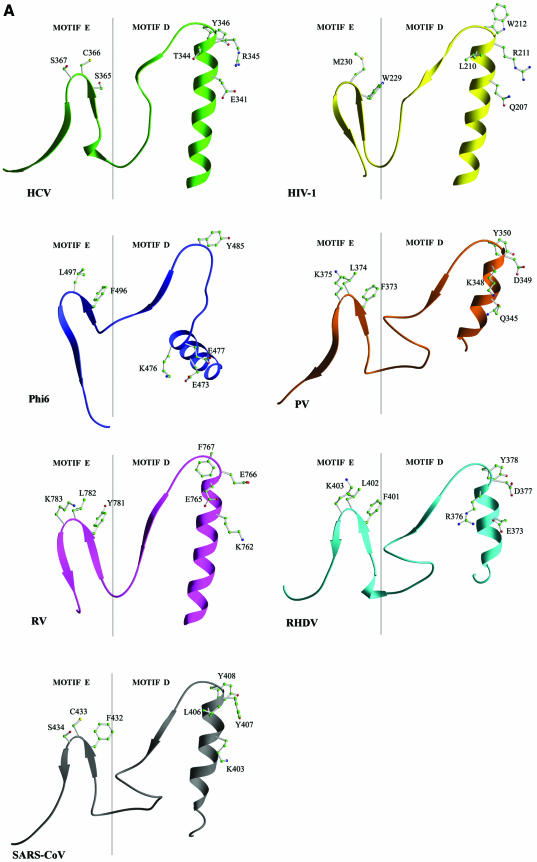
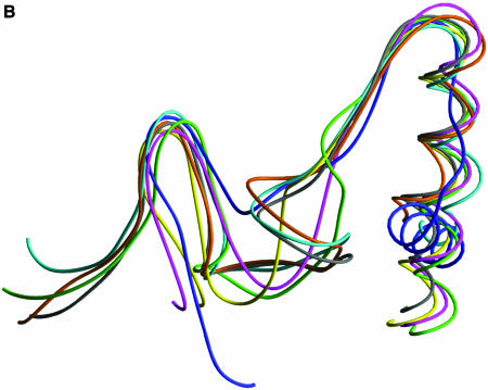
Figure 4.
Structural comparison of HCV, PV, RHDV, RV, φ6 and SARS-CoV RdRps and HIV-1 RT in the regions containing motifs D and E. (A) Ribbon presentation of motifs D and E in the structures of HCV, PV, RHDV, RV, φ6 and HIV-1 polymerases, and in the structural model of SARS-CoV polymerase. (B) Superposition of the regions containing motifs D and E in different viral RdRp and RT structures [the color coding is the same as in (A)].


