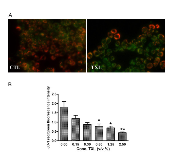
Figure 2.

TXL's effect on the depolarization of HT-29 mitochondria. (A) JC-1 staining observed by fluorescence microscopy. TXL treated cells showed a majority of cells stained green dye due to low mitochondrial membrane potential. (B) Effect of TXL on the depolarization of HT-29 mitochondria was also measured by fluorescence plate reader using JC-1. The cells were exposed to increasing concentrations of TXL. Data represent mean and standard deviation of three individuals with asterisks denoting significant differences between controls and TXL-exposed cells (*P < 0.05, **P < 0.01). CTL: control.
