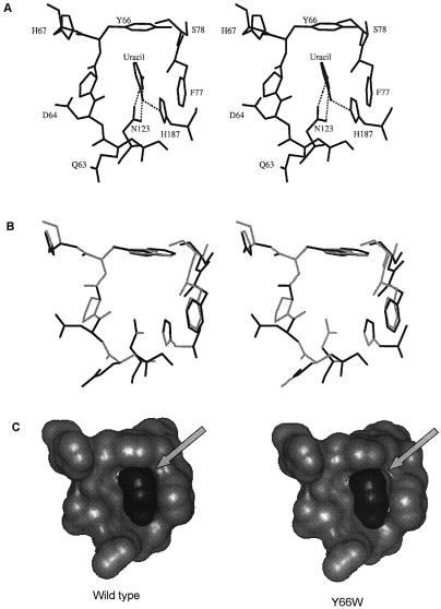
Figure 1.
(A) Uracil specificity pocket showing the interactions between the uracil residue, and the side chains of Y66, F77, N123 and H187 in the active site pocket of E.coli UDG (PDB code 1FLZ). (B) Stereo view of the superposition of uracil specificity pocket of the wild type (light gray) and the Y66W mutant (dark gray) subsequent to removal of short contacts in the Y66W mutant structure. (C) Accessible surface of the pocket (light gray) and the bound uracil (dark gray) illustrating the loosening of the fit in the Y66W mutant. H187 has been removed for clarity.

