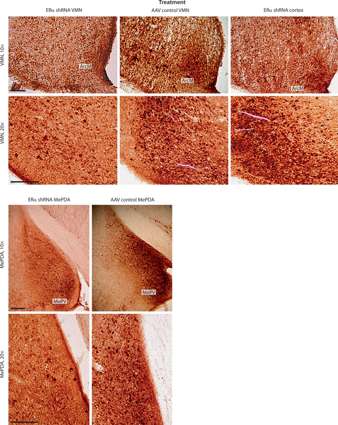
Fig. 2.
Immunocytochemical staining of brain slices in the VMN and the MePDA. Only slices with the greatest ERα staining have been chosen. Scale bar is 200 μm. ArcM = Arcuate nucleus; MePV = medial posteroventral amygdala. Notice a reduction of ERα staining in the VMN and MePDA but not in the ArcM and MePV in animals infused with ERα shRNA compared to animals infused into the cortex and those infused with AAV control.

