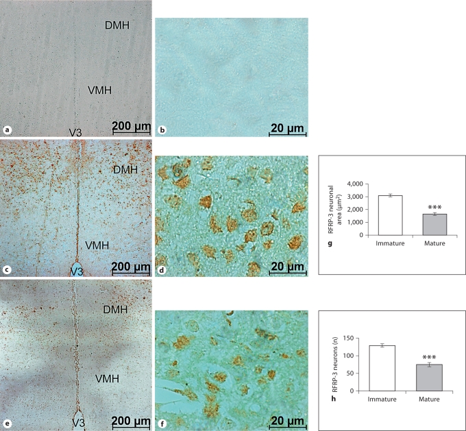
Fig. 2.
Low- and high-magnification coronal sections through the DMH showing the negative control (i.e. not treated with GnIH antiserum; a, b), and the localization of ir-RFRP-3 neurons in the sexually immature (c, d) and mature (e, f) mice. V3 = Third ventricle; VMH = ventromedial hypothalamus; DMH = dorsomedial hypothalamus. The histograms show ir-RFRP-3 neuronal area (g) and the number of ir-RFRP-3 neurons in the DMH (h). Values are means ± SE. ∗∗∗ p <0.001 vs. immature mice.

