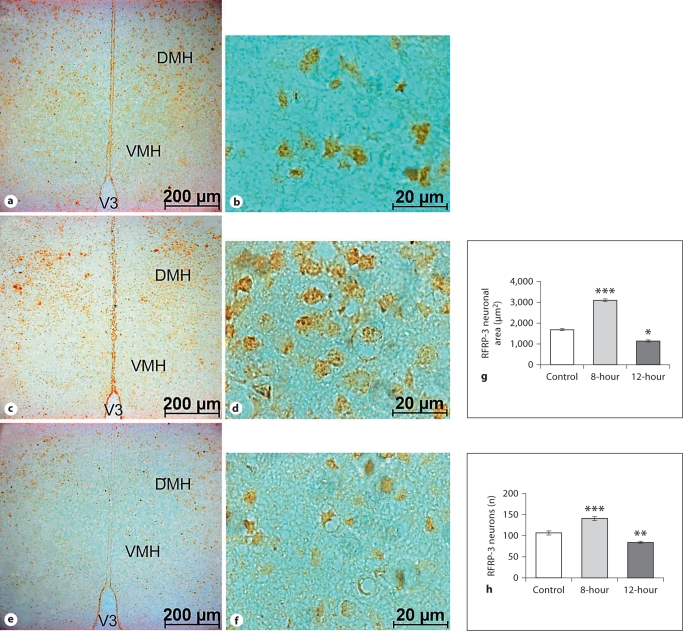
Fig. 6.
Low- and high-magnification coronal sections through the DMH showing ir-RFRP-3 neurons. Compared to the control (a, b), there is an increased number and size of ir-neurons in 8-hour mice (c, d) and a decrease in 12-hour mice (e, f). Increased immunoreactivity is also evident in the RFRP-3 neurons of 8-hour mice, a decrease in the 12-hour mice, as compared to the control. V3 = Third ventricle; VMH = ventromedial hypothalamus; DMH = dorsomedial hypothalamus. Histograms show the RFRP-3 neuronal area (g) and the number of RFRP-3 neurons in the DMH (h). Values are means ± SE. ∗ p < 0.05, ∗∗ p < 0.01, ∗∗∗ p < 0.001 vs. control.

