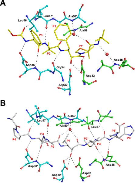
Figure 4.
Hydrogen-bonded interactions between HTLV-1 PR and the inhibitors. A) The binding site of KNI-10562. The inhibitor is shown in yellow sticks, whereas the residues of the enzyme are shown in ball-and-stick representation (molecule A: green, molecule B: cyan). B) Statine-based inhibitor bound to HTLV-1 PR. The inhibitor is shown as gray sticks. Hydrogen bonds are shown in black dashes.

