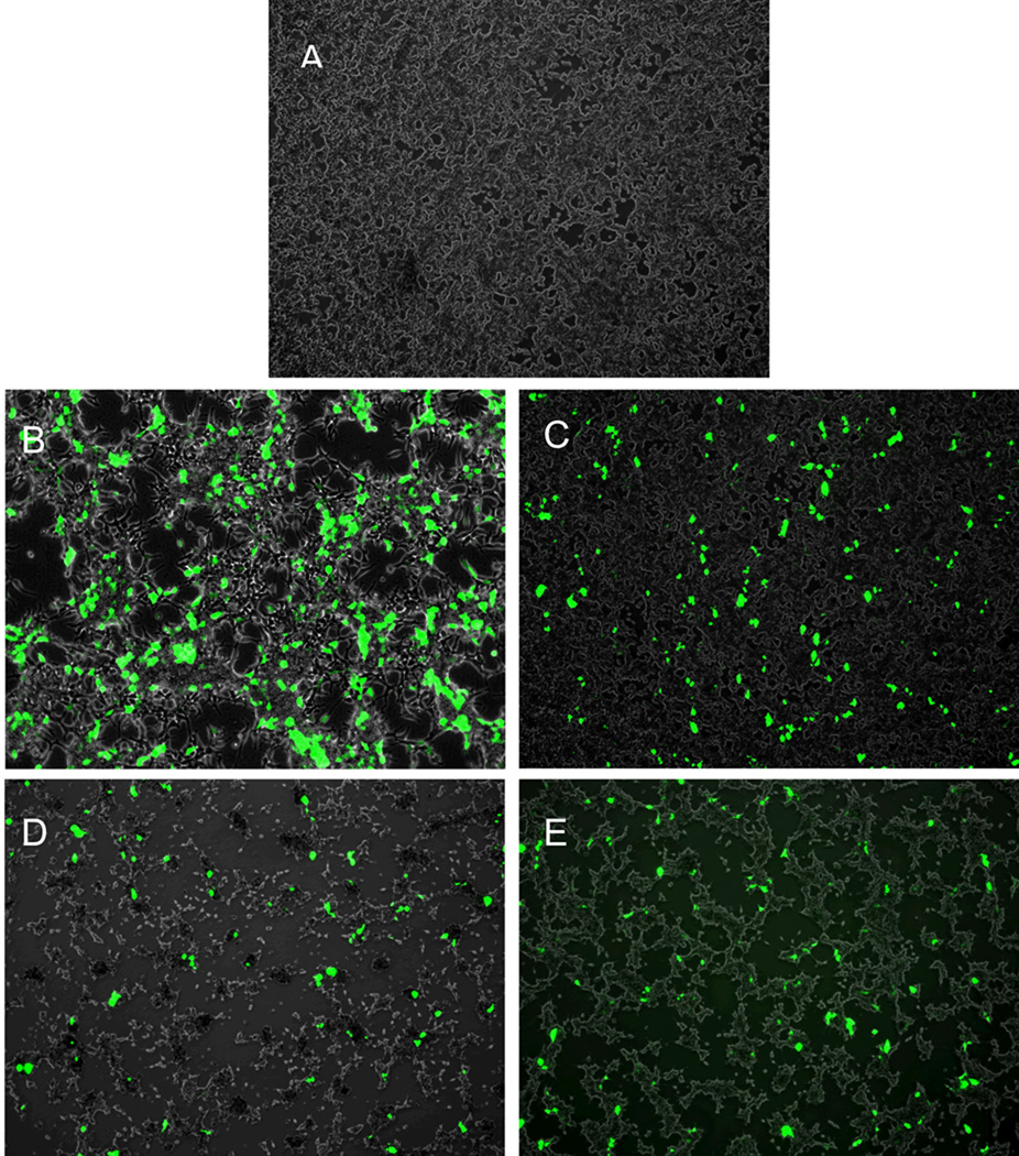
Figure 5.
Fluorescence images of 293T cells (untreated, A) and 293T cells transfected with GFP plasmid mediated with PEI (B), TransIT (C), G4.0 (D), and G4-BAH-PEG42 (E). The cells were exposed to the vector/GFP plasmid polyplexes for 6 hours, rinsed, and then cultured for another 48 hours. Original magnification, ×100.

