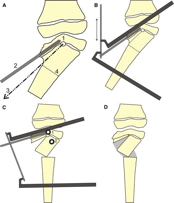
Fig. 5.

Diagrammatic representation of the double elevation osteotomy. a1: antro-posterior tunnel, 2: half pin inserted in the proximal fragment stopping just before the intercondylar eminence, 3: the direction of pull of the Gigli saw, 4: distal osteotomy perpendicular to the tibial shaft anatomical axis. b A two-ring construct assembled, the arrow shows the medial distraction. c After full correction 1: first position of the hinges to achieve correction through the proximal osteotomy, 2: second position of the hinges to achieve correction through the distal osteotomy. Note that this position allows correction of the varus deformity together with lateral translation to align the mechanical axis of the limb
