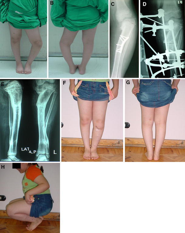
Fig. 6.

Clinical photographs and X-rays of case no. 4, a female patient 9 years old with grade VI infantile tibia vara that had a previous corrective osteotomy followed by recurrence of the deformity. a, b Preoperative clinical photographs, c Preoperative X-ray, d X-ray in the frame, e X-rays obtained 4 years following frame removal, f–h Final clinical photographs showing correction of the deformity and a full range of knee motion
