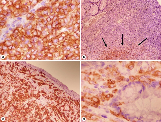
Fig. 2.
a Tumor cells showed strong and diffuse reactivity with C-kit. CD117 stain ×400. b Only 20% of tumor cells showed positivity with HMB-45, in the deeper portion (arrows). HMB-45 stain ×100. c The tumor showed diffuse and strong positivity with Melan-A. Note the absence of junctional features. Mart-1 ×100. d Epithelioid atypical cells infiltrating rectal glands. Mart-1 ×400.

