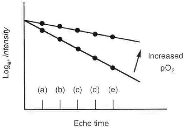
Fig. 1. Correlation of blood oxygenation level-dependent (BOLD) MRI with pO2.

R2* = slope ∼ conc[deoxyHB] ∼ blood pO2 ∼ tissue pO2. The deoxygenation of hemoglobin changes its magnetic characteristics, leading to changes in a parameter of magnetic resonance called R2* (apparent spin-spin relaxation rate). R2* can be estimated from signal intensity measurements made at several different echo times (a through e). The slope of loge (intensity) vs. echo time determines R2* and is directly related to the amount of deoxygenated blood. A decrease in the slope implies an increase in the pO2 of blood. Because blood pO2 is thought to be in rapid equilibrium with tissue pO2, changes in BOLD signal intensity or R2* should reflect changes in the pO2 of the tissue.
