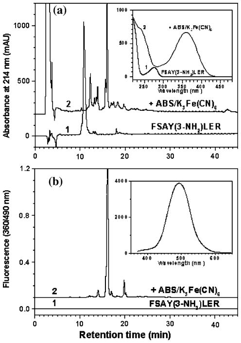
Fig. 3.
Fluorescence tagging of FSAY(3-NH2)LER analyzed by RP–LC with UV detection at 214 nm (a) and by fluorescence detection with excitation and emission wavelengths at 360 and 490 nm, respectively (b). Chromatograms 1 and 2 are recorded before and after tagging of 50 μM FSAY(3-NH2)LER with 10 mM ABS and 0.5 mM K3Fe(CN)6 for 1 h at room temperature. The UV spectra of major peaks analyzed by a PDA detector are shown in the insert to a; the fluorescence spectrum of a total sample is shown in the insert to panel b

