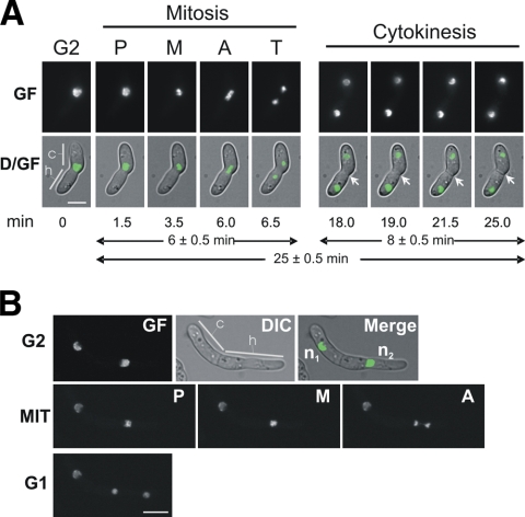
Fig. 2.
Live-cell imaging of mitoses after conidial germination. (A) First nuclear division in a germling of a FoH1::GFP strain. Images were recorded consecutively for GF and Nomarski optics (DIC) (see Materials and Methods). D/GF, merged images for DIC and GF. Conidial (c) and hyphal (h) compartments are indicated. Mitotic phases are indicated as follows: P, prophase, M, metaphase, A anaphase, and T, telophase. After karyokinesis, the formation of a septum was visualized (arrows). Scale bar, 5 μm. The time in minutes at which each micrograph was taken (see Movie S2 in the supplemental material) and the average times for mitosis and cytokinesis are indicated below the images. (B) Second nuclear division in a FoH1::GFP strain. The upper row shows fluorescence (GF), DIC, and merged images of a cell with two compartments at G2 stage. Nuclei are indicated as n1 and n2. Note that while n1 remains interphasic, n2 undergoes mitosis. Samples were grown and imaged as described for panel A. Mitotic phases (MIT) and cell type are indicated. Scale bar, 5 μm.

