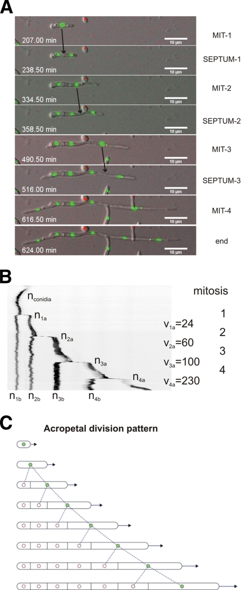
Fig. 3.
Patterns of nuclear division in F. oxysporum. (A) Series of images of a developing hypha from Movie S3 in the supplemental material showing 4 consecutive mitoses, each followed by formation of a septum. The time in minutes (seconds in decimal scale) is documented in Movie S3 in the supplemental material. The images were acquired using an Axioplan2 microscope equipped with appropriate filters and a CoolSnap HQ camera at room temperature (RT) every 90 s for 15 h simultaneously for GFP and ChFP fluorescence and Nomarski optics, and merged for DIC and green and red fluorescence. (B) Kymograph of nuclear fluorescence (inverted image), using the Metamorph software package, from a selected hypha. Note that mitosis occurs exclusively in the apical compartment of a vegetatively growing hypha, generating one mitotically active daughter nucleus (shown in green in the diagram in panel C) and one mitotically dormant daughter nucleus (in white in the diagram in panel C). The mitotically active nucleus is located in the apical compartment (indicated as nxb, where x is the mitosis number) and the dormant nucleus in the subapical compartment (indicated as nxa, where x is the mitosis number). The speed of the apical nucleus after mitosis and apical-compartment elongation (vx) is indicated in nm min−1. (C) Model of the process. Filled circles, active nuclei; empty circles, dormant nuclei.

