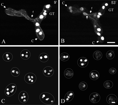
Fig. 1.
Timing of fluorescent protein labeling of heterokaryons formed by CAT fusion. (A and B) Fused conidial germlings (one labeled with H1-GFP and the other labeled with β-tubulin-GFP [BML-GFP]), one 9 min (A) and the other 53 min (B) after fusion. At the 9-min time point, BML-GFP has become incorporated into microtubules derived from the left-hand germling with nuclei carrying the hH1-sgfp gene, while HI-GFP has not yet labeled the nuclei in the germling on the right-hand side with nuclei carrying the bml-sgfp gene. Microtubules passed through the fused CATs (f) before nuclei did. The location of the spindle pole body (arrow) with associated microtubules is shown at 53 min. C, conidium; GT, germ tube. Bar, 10 μm. (C) Conidia from a homokaryotic H1-GFP strain. Each conidium has several nuclei. (D) Conidia from a heterokaryotic H1-GFP plus β-tubulin-GFP strain visualized by confocal microscopy. Variable levels of GFP expression were observed for different conidia. Bar, 5 μm.

