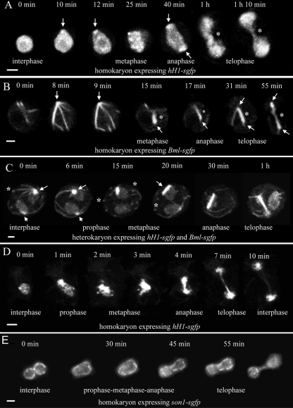
Fig. 2.
Time courses of mitosis in multinucleate nongerminated conidia (A to C) and germ tubes (D and E). The interphase, prophase, metaphase, anaphase, and telophase stages of mitosis have been indicated where they can be clearly recognized. (A) Mitosis of a conidial nucleus with H1-GFP labeling. A bright fluorescent nuclear region (arrows) was often evident where a spindle pole body (SPB) should be located. The segregating H1-containing chromatin was connected by a “chromatin bridge” (*) from late anaphase to telophase. Bar, 2 μm. (B) Mitosis of a conidial nucleus labeled with β-tubulin-GFP. Microtubules were formed from a focal point presumed to be the SPB (arrows). During late prophase/early metaphase, microtubules bundled to form a compact spindle (*), which is evident from metaphase to telophase. Bar, 2 μm. (C) Mitosis in a heterokaryotic conidium in which two nuclei labeled with H1-GFP and β-tubulin-GFP were dividing asynchronously (only one nucleus divided during this time course). Astral microtubules are formed from the single SPBs (arrows) in nuclei during both interphase and early prophase. These microtubules often possessed a bright focal point where they were in contact with the plasma membrane (*). SPB duplication occurred during prophase (at 15 min). The spindle developed as a compact bundle of microtubules (15 min to 1 h). During this time, H1-GFP was associated with the spindle and by telophase had segregated to both SPBs (the chromatin bridge is masked by the BML-GFP in this image). Astral ray microtubules can be visualized at the 1-h time point. Bar, 4 μm. (D) Mitosis in a germ tube nucleus labeled with H1-GFP. In this sequence, the expression level of H1-GFP was much lower than that in C. Bar, 2 μm. (E) Germ tube nucleus labeled with SON-1-GFP to show the nuclear pores in the nuclear envelope. Telophase was easily observed because the bridge formed between the nuclei daughters. Projections of 12 deconvolved images are shown. Bar, 2 μm.

