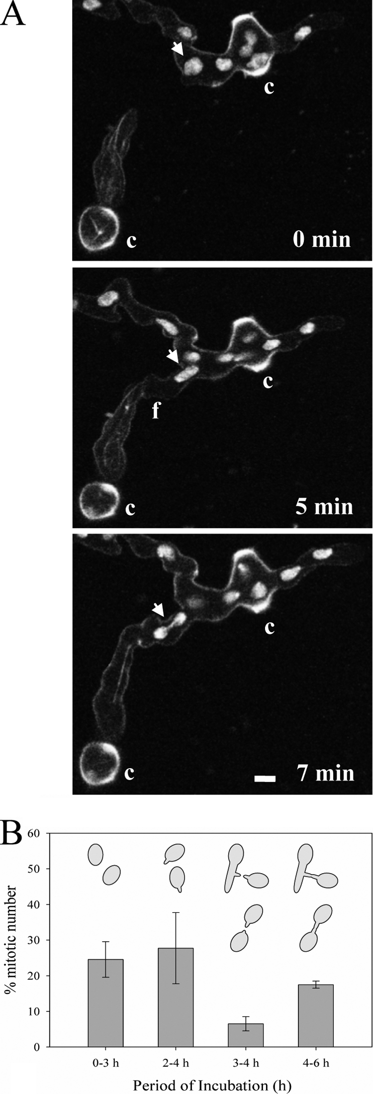
Fig. 4.

Cell cycle arrest during CAT homing. (A) CATs formed from germ tubes before (0 min) and after (5 and 7 min) fusion visualized by confocal microscopy. The upper conidial germling was labeled with H1-GFP, while the lower germling was labeled with β-tubulin-GFP. Bar, 5 μm. (B) Percentages of nuclei undergoing mitosis in ungerminated macroconidia (0 to 3 h), in macroconidia possessing only germ tubes (2 to 4 h), in macroconidia or macroconidial germlings possessing CATs (3 to 4 h), and in macroconidia or macroconidial germlings that have undergone CAT fusion (4 to 6 h). The time periods selected for the analysis of mitosis in these four different cell types were those in which the production of the individual cell types was maximal. Mitotic nuclei were identified by the presence of thick microtubule bundles in spindles labeled with BML-GFP.
