Fig. 7.
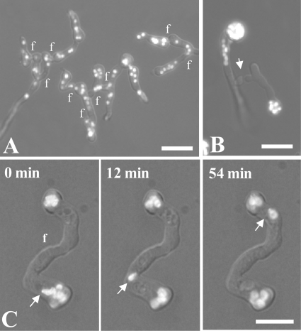
Nuclear distribution and behavior in the wild type and ropy (dynein/dynactin) mutants visualized by combined wide-field, bright-field, and fluorescence microscopy. Nuclei were labeled with H1-GFP. (A) Network of fused wild-type germlings showing a more-or-less random distribution of nuclei. (B) ro-1 (dynein heavy chain) mutant showing CATs derived from germ tubes homing toward each other (arrow). The nuclei are concentrated in or close to the conidia. (C) Δro-11 (dynactin subunit) mutant showing nuclear migration (arrow) through fused CATs (f) after incubation for 7 h. f, points of CAT fusion. Bars, 15 μm.

