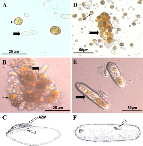
FIG. 1.
Micrographs and schematic drawings of the two ciliates detected in this study. (A to C) Samples (Cil1) from Acropora microclados before (A) and after (B) culturing and drawing (C) to show the peristome (arrowhead), adoral zone of membranelles (AZM) (straight arrow), and feeding current (curvy arrows) generated by the vibration of AZM. (D to F) Samples (Cil2) from Acropora hyacinthus before (C) and after (D) culturing and drawing (F) to show the peristome (arrowhead). Thick filled arrows denote the dominant ciliates; thin filled arrows denote Symbiodinium.

