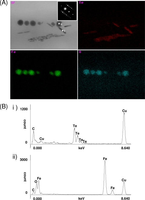
FIG. 4.
STEM-EDX and SAED analyses for magnetite and tellurium within magnetotactic bacteria. (A) Bright-field (BF) STEM image with SAED patterns of a rod-shaped crystal and STEM-EDX maps of Te, Fe, and O taken using a probe size of approximately 2 nm; (B) spot scans of *i and *ii as representations of tellurium and magnetite, respectively.

