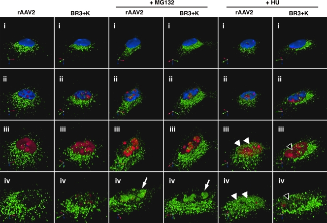
FIG. 7.
HU treatment does not mobilize BR3+K capsids to the nucleoplasm. To visualize whether BR3+K mutant capsids could be mobilized into the nucleoplasm in response to HU, we fixed and stained infected HeLa cells for indirect immunofluorescence of capsids (green), nucleolin (red), and nuclei (blue). Roughly 40 confocal sections that span 5 μm were stacked and rendered in three dimensions by using Volocity software (i). A movie was generated to explore capsid localization in detail by digitally gating first the blue channel (DAPI, representing the nucleus) and then the red channel (nucleolin, representing the nucleolus), along with increasing the zoom. Images were captured at successive stages in the movie to reveal the nuclear interior (ii and iii) and the interior of the nucleolus (iv). Both rAAV2 and the BR3+K mutant accumulate in the nucleolus in the presence of proteasome inhibitor (white arrows). Only rAAV2 is localized to nucleoplasmic sites following HU treatment (white arrowheads, rAAV2; empty arrowheads, BR3+K mutant).

