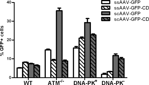
FIG. 5.
ssAAV and scAAV circularization assay with DNA-PKCS-deficient cells. Cells were infected with the four reporter vectors (see Fig. 1) at an MOI of 1 for 2 h. GFP expression was quantified by flow cytometry at 24 h postinfection. Means decreases for transduction in DNA-PK+ and DNA-PK− cells for each virus are all significantly different (P < 0.0001 for ssAAV-GFP, ssAAV-GFP-CD, and scAAV-GFP-CD; P = 0.0002 for scAAV-GFP). Error bars indicate standard deviations.

