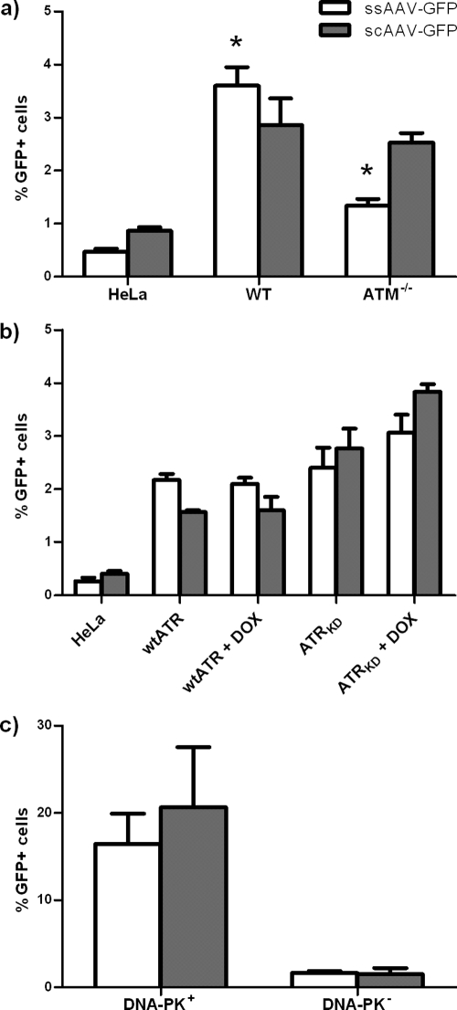
FIG. 8.
Integration of ssAAV-GFP and scAAV-GFP vectors in ATR-, ATM-, and DNA-PKCS-deficient cells. (a) wt and ATM−/− cells were infected at an MOI of 1,400 for 2 h. Cells were passed and GFP expression was quantified by flow cytometry until the percentage of GFP-positive cells reached a plateau at 22 days postinfection. The indicated (*) differences between wt and ATM-deficient cells were significant (P < 0.0038). (b) Doxycycline-treated and untreated wt ATR and ATRKD cells were infected for 2 h at an MOI of 260 Treatment with DOX was maintained until 2 days postinfection. Cells were passed and GFP expression was quantified by flow cytometry until the percentage of GFP-positive cells reached a plateau at 25 days postinfection. Means for percent GFP-positive cells for ssAAV-GFP and scAAV-GFP vectors were not significantly different for wt ATR and wt ATR + DOX cells or for ATRKD and ATRKD + DOX cells. (c) DNA-PK+ and DNA-PK− cells were infected at an MOI of 1,400 for 2 h. Cells were passed and GFP expression was quantified by flow cytometry until percentage of GFP-positive cells reached a plateau at 48 days postinfection. Means for percent GFP-positive cells for ssAAV-GFP and scAAV-GFP vectors are significantly different (P = 0.0018 and P = 0.0088, respectively). Error bars indicate standard deviations.

