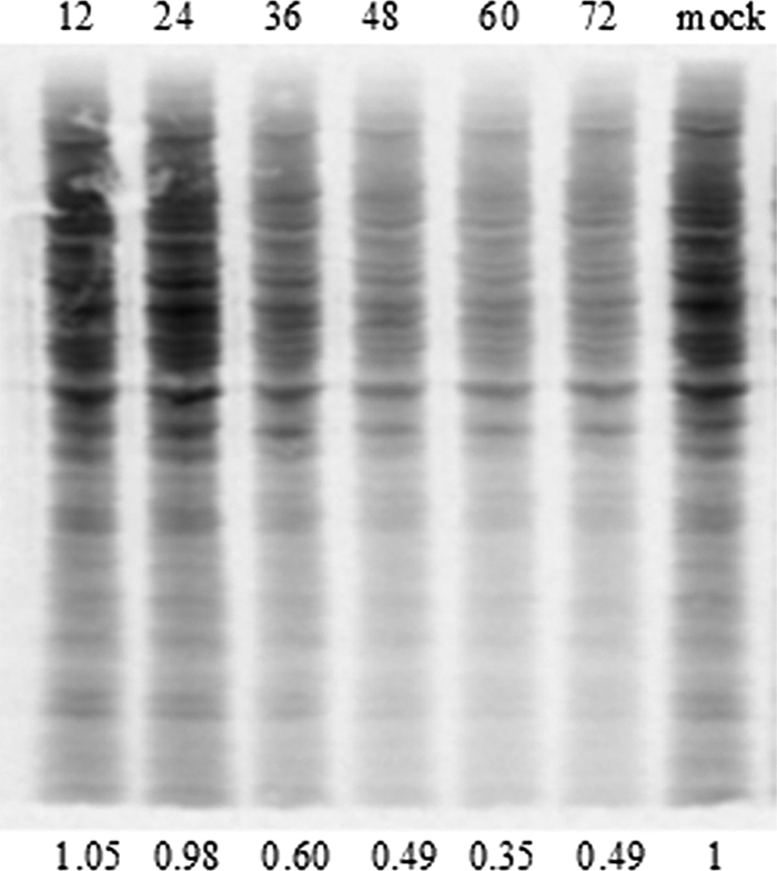
FIG. 10.

Residual protein synthesis in TO cells infected with SAV-3. Confluent TO cells were grown in a six-well plate and infected as described in Materials and Methods. The membrane was exposed in a PhosphorImager cassette and then scanned by using a Typhoon imager. Numbers above the figure represent hours postpulsing. The protein amount was quantified by densitometry with ImageJ software as described in Materials and Methods. The results show 40, 51, and 65% reduction by 36, 48, and 60 h postinfection, respectively. Mock infection is shown at the far right.
