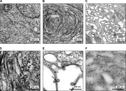
FIG. 8.
Ultrathin-section EM of infected cells. 293T cells were infected with TBEV strain Hypr (MOI of 5) for 24 h before fixation and processing for regular transmission EM (A to E) or immuno-EM (F). (A) Transmission EM of mock-infected cells. (B) Transmission EM of TBEV-infected cells. (C) Transmission EM of TBEV-infected cells treated with BFA at 12 h p.i. (D and E) Close-up EM pictures of untreated and BFA-treated cells that were infected with TBEV. (F) Close-up immuno-EM picture of cells infected with TBEV. Immunogold staining was performed by using anti-dsRNA mouse monoclonal J2 as the primary antibody and goat anti-mouse IgG/IgM conjugated with 10-nm colloidal gold as the secondary antibody. Arrowheads indicate virus particles, lined arrows indicate virus-induced membrane vesicles, and the boxed arrow indicates immunogold-labeled dsRNA.

