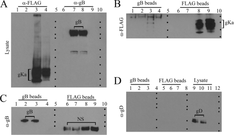
FIG. 2.
The gKa peptide interacts with gB. Vero cell monolayers were mock transfected, transfected with a plasmid expressing the gKa peptide under the control of the HCMV-IE promoter, or a control plasmid containing the gB coding sequence in a reverse orientation. Twenty-four hours posttransfection, cells were infected with HSV-1(F) gKΔ31-68 virus, and after 24 hpi, cellular extracts were prepared and examined for the presence of gB or gKa before and after immunoprecipitation with either anti-gB or anti-FLAG (anti-gKa). (A) Lanes 1 to 4 show immunoblots reacted with anti-gKa antibody. Lane 1, mock-transfected cell extracts; lane 2, cellular extracts transfected with the control plasmid and subsequently infected with gKΔ31-68 virus; lane 3, cellular extracts transfected with the gKa plasmid and subsequently infected with gKΔ31-68; lane 4, gKa-transfected cellular extracts. Lanes 6 to 9 represent the same samples as those in lanes 1 to 4, respectively, except that the immunoblot was probed with anti-gB antibody. Lanes 5 and 10 demarcate the relative positions of the molecular mass markers (10 to 250 kDa). (B) The sample arrangement in of immunoblot is the same as that described above (A) except that all cellular extracts were first immunoprecipitated with either anti-gB (lanes 1 to 4) or anti-gKa (lanes 6 to 9) antibody and the immunoblot was probed with anti-gKa antibody. Lanes 5 and 10 demarcate the relative positions of the molecular mass markers (10 to 25 kDa). (C) The sample arrangement of the immunoblot is the same as that described above (B) except that the immunoblot was probed with anti-gB antibody. Lanes 5 and 10 demarcate the relative positions of the molecular mass markers (75 to 250 kDa). (D) Immunoblot probed with anti-gD antibody. Lane 1, cellular extracts transfected with control plasmid and subsequently infected with the gKΔ31-68 virus; lane 2, cellular extracts transfected with the gKa plasmid and subsequently infected with gKΔ31-68; lane 3, gKa-transfected cellular extracts. Samples represented in lanes 1, 2, and 3 were immunoprecipitated with anti-gB antibody and probed with anti-gD. Lanes 5 to 7 are the same as lanes 1 to 3, respectively, with the exception that samples were immunoprecipitated with the anti-FLAG antibody and probed with anti-gD antibody. Similarly, lanes 9 to 11 are lanes 1 to 3, respectively, probed with anti-gD antibody without any prior immunoprecipitation. Lanes 4, 8, and 12 demarcate the relative positions of the molecular mass markers (37 to 250 kDa).

