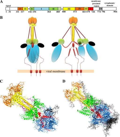
FIG. 8.
Schematic representation of potential interactions between gK and the three-dimensional X-ray structure of gB derived by protein docking algorithms. (A) Linear domain architecture of gB as previously shown by Heldwein et al. (23). (B) Schematic representation showing gKa interacting with gB domains I and II (23). (C) Schematic representation of the top-ranked gKa-gB complex produced by the ZDOCK and ZRANK algorithms. (D) Schematic representation of 15 top-ranked positions where gKa may interact with gB.

