
Figure 3.
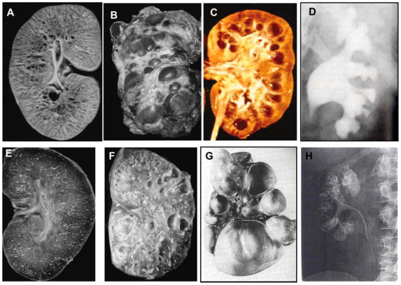
Kidney disease in ciliopathies. A: Kidneys of an infant with ARPKD that are enlarged with preserved contours due to uniform microcystic non-obstructive dilatation of collecting ducts. B: Massivelyenlarged kidneys of an adult with ADPKD that display large macrocysts distorting the kidney contour. C: Kidneys of an individual who has renal failure due nephronophithisis. D: Intraveneous pyelography of Bardet–Biedl syndrome kidneys displaying clubbing, blunting, and distortion of renal calyces in the absence of distal obstruction. E: Kidneys of an individual with glomerulocystic kidney disease displaying small cysts mostly located in the renal cortex. F: Cystic dysplastic kidneys in an infant with Meckel–Gruber syndrome. G: Multicystic dysplastic kidneys. H: Intraveneous pyelography displaying medullary sponge kidney. Sizes of the kidneys are not to scale. (A, B, E, and F are from Bisceglia et al., 2006, C is from Hildebrandt and Zhou, 2007, D is from Alton DJ and McDonald MB, Radiology, 109: 659–663, 1973. G is from Zerres et al., 1984. H is from Heptinstall’s Pathology of the kidney, Ed: Jennette J., Olson J., Schwartz M., Silva F.)