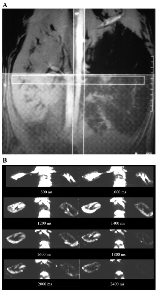
Fig. 1.

Renal perfusion magnetic resonance imagery (MRI) by arterial spin labeling (ASL). A: method of acquiring MRI following ASL. The vertical stripe illustrates the labeling radio frequency (RF) pulse applied so that all the spins in the descending aorta are inverted. The horizontal stripe is a representation of the transverse slice of interest through the kidneys. The thick hatched slab (right) represents a presaturation pulse applied to avoid any venous contribution to the observed signal intensity and to minimize any artifacts due to peristaltic motion in the abdomen. Two acquisitions were performed, one with and one without the labeling pulse. The difference image would then be a representation of all the labeled blood that enters the slice of interest. Obviously, this varies as a function of time delay between the labeling pulse and the acquisition. B: set of transverse (difference) images through the kidneys obtained after different delay times in a swine model with a chronic renal artery stenosis in the left kidney (right). Based on this set of data, it is possible to estimate absolute perfusion (115).
